TIPS & TRICKS - Incision of a Stenotic Calyceal Infundibulum
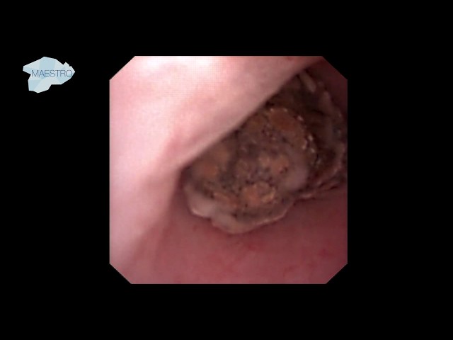
A URS for a symptomatic 2 cm renal stone in the lower pole is performed here. The stone was initially not found during ureteroscopy but was clearly visible in the CT scan. During the exploration of the intrarenal cavities, a small opening was identified on the lateral surface of the renal pelvis. Identification of the stenotic calyceal infundibulum in the lateral wall of the renal pelvis of a left kidney. A small incision at 3 o’clock of the stenotic calyceal infundibulum was made. Urine coming out of the calyx is visible at this point. The incision was extended laterally to enlarge the neck of the infundibulum until the stone appeared. In the following step the infundibulum was enlarged to let the scope enter the calyx and chase the stone properly.